
Tags
biology, biotechnology, DNA, DNA nanotechnology, drug delivery, Nanotechnology, origami, Popular science, science, Science fiction, shape, tetrahedron
 In the previous post, I introduced DNA nanotechnology and talked a bit about some of the 2-D structures that researchers have devised. Scientists have also used these techniques to build a variety of 3-D shapes, some of which can be used as containers for drug delivery. In this post, I’m going to focus on the DNA tetrahedron, a four-sided pyramid made of DNA which is relatively easy to build and manipulate and has proven useful in several recent studies.
 One way of building a DNA tetrahedron is to combine four subunits (pictured left), each of which folds to become one corner of the pyramid. The subunits are made by stitching together seven strands of DNA for a total of 28 strands in the entire structure. The resulting pyramid is about 10 nanometers tall — a nanometer is a billionth of a meter, so you could stack around 10,000 DNA tetrahedrons in the width of a human hair.
Last year, a group of researchers figured out a way to add and remove a cap to the faces of the pyramid, effectively sealing and unsealing the cage. They added short stretches of DNA to each subunit (shown in purple above); these extra bits don’t become part of the pyramid’s structure but instead dangle from the edges, hanging into the space between them and acting as anchors to which the cap can attach. Each cap is made of a single long strand of DNA bent into a triangle with three shorter strands attached to cover most of its length, leaving a few regions exposed to connect to the anchors on the pyramid. When the caps and pyramids are mixed together, the anchors bind to the parts of the cap that aren’t blocked by short strands and the cap covers the face of the pyramid. The caps can be removed by adding specially designed DNA strands that are better than the short strands and the anchors at connecting to the long DNA strand in the cap; they bind to the cap, knocking the small strands and anchors out of place and releasing the cap from the pyramid. It’s a remarkable feat of bioengineering that’s hard to capture in words, so I’ll fall back on the researchers’ figures for some help:
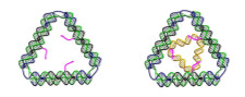
A single face of the pyramid without a cap (left) & with a cap (right). (Image modified from Zhang et al.)
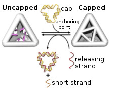
The capping & uncapping process. (Image modified from Zhang et al.)
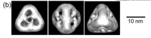
An image of the capped DNA tetrahedron based on electron microscopy. (Image adapted from Zhang et al.)
Although the researchers figured out how to seal and unseal a DNA pyramid nanocage, they didn’t go as far as putting something in it — but that’s how science usually proceeds, step by careful step. Another team of scientists has used the DNA tetrahedron to carry drugs into cells, but they took a somewhat different approach. They wanted to find out how well DNA tetrahedrons could deliver the anti-cancer drug doxorubicin into breast cancer cells, and whether this could help to overcome drug-resistance in some of the cells. The drug can slide into the space between the A, T, G, and C that make up DNA, so it integrated directly into the edges of the pyramid, removing the need for capping and uncapping in this case. Normal breast cancer cells took up doxorubicin equally well regardless of how it was delivered; resistant cells, on the other hand, only accumulated doxorubicin delivered in a DNA tetrahedron. These cells pump free doxorubicin out of themselves to escape its effect, but embedding the drug in a DNA tetrahedron somehow stops them from being able to do that. The researchers think this might be because the tetrahedron enters the cell in a different way than the drug and ends up protected inside an endosome. The conditions within the endosome make the tetrahedron break apart, releasing the drug into the cell. Although there’s still a lot more work to be done, this experiment hints at some of the exciting promise of DNA nanotechnology.
DNA nanotechnology is a relatively young and rapidly changing field. I’m not an expert in it and these two posts certainly aren’t supposed to be a comprehensive overview — they’re just a summary of a few interesting experiments and techniques. I think this is going to be a pretty active area of research and hopefully one that will yield impressive fruit as the technology matures. I’ll certainly be keeping an eye on it and will probably revisit the subject once I’ve learned more!
Refs
Zhang C, Tian C, Li X, Qian H, Hao C, Jiang W, & Mao C (2012). Reversibly switching the surface porosity of a DNA tetrahedron. Journal of the American Chemical Society, 134 (29), 11998-2001 PMID: 22800434
Kim KR, Kim DR, Lee T, Yhee JY, Kim BS, Kwon IC, & Ahn DR (2013). Drug delivery by a self-assembled DNA tetrahedron for overcoming drug resistance in breast cancer cells. Chemical communications (Cambridge, England), 49 (20), 2010-2 PMID: 23380739

This is certainly very interesting, thank you for sharing! Receptors on the surface of cells, aka cell receptivity, is the current methodology for introducing drugs into cells to produce the therapeutic effect ie: acetaminophin for ‘numbing’ pain receptors. This tetrahedron on the DNA level is very fascinating, I’m definitely intrigued, potentially concerned, as to where this research will lead us.
Reblogged this on mistyjdjrx.